Ferret Anemia
- May 11, 2017
- Posted by: Santiago Diaz
- Categories: Office, Patients
Ferrets are very social, playful and make excellent pets. Nevertheless, like any other animal, they are prone to certain diseases in captivity. Adrenal disease is probably the most common disease observed in ferrets in the United States. Today, we are going to cover a case of a ferret that had severe anemia.
Anemia is a condition characterized by a deficiency of red blood cells (erythrocytes) in the blood. Red blood cells transport oxygen to all the tissues in our body, which is then utilized by cells for normal function and metabolism. Red blood cells transport oxygen by binding it to hemoglobin, an iron-containing complex protein.
Anemia can be seen with different conditions including active bleeding, bone marrow abnormalities, immune-mediated diseases, iron deficiency, chronic inflammatory disease, and other disease processes.
Anemia can be identified by measuring the packed cell volume (PCV) or percentage of red blood cells in circulation. A normal PCV for ferrets is 43-55%.
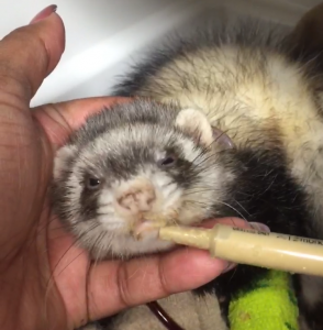
The following ferret is a 3 year old spayed female ferret that was referred to us by a local veterinarian for a blood transfusion. She had a PCV (packed cell volume) of 9%, her mucous membranes were white and tacky, and she was barely responsive. Her body temperature was 96.9 F. Patient had a grade I/VI systolic heart murmur as a result of the anemia. She had a severe infestation of fleas that were draining her of her blood.
We immediately contacted one of our clients that has healthy ferrets, and she graciously volunteered one of her biggest ferrets to donate blood for a transfusion. We picked up her ferret, performed an exam and after it was determined he was healthy and had a normal PCV, we obtained 12 ml of whole blood. We mixed the blood with ACD solution, which is a blood preservative and anti-coagulant solution.
The anemic ferret was then pre-medicated with diphenhydramine (Benadryl) to prevent any allergic reactions from the transfusion and shortly thereafter the transfusion was started. For the first 30 minutes all of her vitals were recorded. After this period, her vitals were recorded every 30 minutes.
We manually removed all the fleas and gave her a dose of selamectin (Revolution) to treat for fleas. We also gave her an iron injection, so that the bone marrow could utilize the iron to create more red blood cells. After the first hour, she was more responsive and took 3 mL of oxbow carnivore care diet. We continued feeding her small amounts of this diet every 1-2 hours
Her PCV was measured 24 hours after the transfusion was started and the result was a PCV of 17%. She also passed stool for the first time after 28 hours. Once the blood transfusion was completed, patient was discharged to continue supportive care at home.
Owner brought her back in one week to recheck her PCV, which was 18%. A second iron injection was given and it was determined that the patient had gained weight and was more bright, alert and responsive. She was still not eating on her own, but was taking the oxbow carnivore care well.
Two weeks after initial presentation, the patient was acting completely normal and her PCV had gone up to 27%. She was eating on her own and had no fleas. A month later, her PCV was completely normal. She had become very active, playful and had a great appetite.


